The Diagnostic Ultra-portable X-ray for Space (DUXS) program achieved liftoff late last year by successfully obtaining medical diagnostic radiographic images in zero-gravity (Zero-G) conditions – including a re-creation of one of the first X-ray images ever taken by Nobel Prize winner Wilhelm Conrad Röntgen. The purpose of this study was to prove successful X-ray imaging is possible in a zero-gravity environment and show that existing equipment is capable of producing diagnostic images in both zero-gravity and Low Earth Orbit (LEO).
The DUXS program is a partnership between industry and academia aiming to improve medical diagnostic capabilities in the final frontier and beyond. True weightlessness, which was required for this study, was accomplished by reserving space in a multi-parabolic flight provided by Zero Gravity Corporation (Zero-G) in G-FORCE ONE, a specially modified Boeing 727. During the flight, highly trained pilots perform a series of parabolas, between 24,000 and 32,000 feet which provides 20 to 30 seconds of Zero-G per parabola.
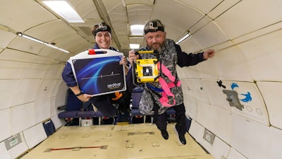
This flight's imaging team consisted of four members. Sheyna Gifford, MD, MPH, MBA, MS focuses her research on preventative medicine, rehabilitation and parastronautics. David Lerner, MD is a researcher from the University of Washington specializing in radiology, as well as remote and aerospace medicine. These two researchers partnered with MinXray Inc., a leading global supplier of portable, compact digital imaging equipment. Vice President of Marketing and Sales, Jeanne Walter, supplied MinXray's Impact Battery-Powered Portable Digital Radiography System, and Director of Global Sales, Mike Cairnie, took the images during the Zero-G flight. These images included a radiographic image of Dr. Gifford's hand, which mirrored the first X-ray ever taken of Röntgen's wife's hand back in 1895 and provided the first conclusive evidence that medical-quality diagnostic X-rays can be quickly and simply obtained in microgravity.
Successful acquisition of radiographic images mid-flight helped identify the complexities that exist when imaging in Zero-G conditions and potential solutions for those problems. Establishing the single inertial frame between all components and participants, which is required for the system to capture quality diagnostic images, was a variable examined by the team during the pre-flight preparation. Several methods of stabilizing the X-ray generator, patient and digital detector together were developed and even modified during the actual flight as the testing progressed.
A line pair phantom was used to establish image quality compared to images acquired in normal earth gravity. Additional images of a patient's hand, elbow, knee and thorax were acquired. The equipment functioned perfectly in the zero-gravity environment with diagnostic quality radiographs acquired comparable to those obtained in normal earth gravity.
Bringing this technology to space has several benefits. X-ray imaging provides additional diagnostic capabilities apart from ultrasound, which is currently used on NASA flights. Successful acquisition of diagnostic radiographic images is possible with minimal training required for the operator. Image acquisition is quick, and images, if required, are easily transmitted for diagnosis. Rapid diagnosis of numerous medical conditions and acute trauma such as fractures, dislocations, respiratory issues or obstructions is now achievable in a Zero-G or LEO environment.
The usefulness of X-ray capabilities in space can be calculated by referencing an IMM Medical Conditions list compiled by NASA. The list cites 100 medical conditions, 47 of which have occurred during a flight and 53 considered possible, and 54 imageable diagnoses. Ultrasound or X-ray alone can diagnose 46 of the 54. However, when both medical imaging modalities are available, 53 of the 54 imageable conditions can be diagnosed. In addition, an X-ray system can be easily integrated into existing diagnostic technology with X-ray and ultrasound images able to be acquired onto the same laptop. An X-ray can also be used for non-destructive testing to possibly assist in maintaining the health of the vehicle, station or other equipment on board without the need for dismantling.
X-ray technology has not yet been utilized for space travel because historically, X-ray equipment has been too large and cumbersome to be considered. However, the MinXray Impact Wireless system used on the Zero-G flight defies these limitations, consisting of MinXray's TR90BH battery powered portable X-ray generator, wireless CsI digital detector and system laptop with the MinXView imaging software package. The Impact system weighs only 31 lbs., is compact, extremely user friendly, produces high quality radiographic images, can integrate with existing diagnostic setups and features off-the-shelf availability with no installation required.
This flight is part of MinXray's ongoing efforts to make diagnostic imaging accessible to everyone regardless of environmental conditions or location. Cairnie has taken this mission around the world, bringing MinXray imaging systems to countries with limited access to medical resources. He has traveled to numerous countries including Nigeria, Uganda, Mongolia and Nepal to screen for tuberculosis in communities highly impacted by the disease. Recently, Cairnie brought X-ray technology to record heights with a mission to the base camp at Mount Everest, making important diagnostic information available, even at extreme elevations.


